The size of catheters: an important parameter to
consider in assessing infectivity
Type of article: Mini review
Sidi Mohammed Lahbib Seddiki*1,2, Zahia Boucherit-Otmani2
1: University Center of Naâma, Algeria
2: Laboratory: Antifungal Antibiotic, Physico-Chemical
Synthesis and Biological Activity, University of Tlemcen, Algeria
Abstract
The diagnosis of catheters’ infectivity is established
considering several parameters. These relate to the clinical patients’ data and
the microbial load of the catheters following their culture. Catheter
infectivity type is related to the significance threshold. However, differences
in sizes exist between several catheters. So, in order to qualify any microbial
alteration, it is important to take into account the impact of the size of the
catheters. For this, future studies should consider this parameter to assess
microbial load properly.
Key words: Catheters; Infectivity; Diagnosis;
Microbial Colony Forming Unit.
Corresponding author:Sidi Mohammed Lahbib Seddiki,University Center of Naâma,
Algeriaseddiki.med@gmail.com
Received: 1 February,
2019, Accepted: 16 Mars, 2019, English editing: 16 Mars, 2019, Published: 01
April, 2019.
Screened by
iThenticate..©2017-2019 KNOWLEDGE KINGDOM PUBLISHING.
1. Important concepts
The ascertainment of this article is the result of
several published studies. Despite notable progress in the study of microbial
infectivity of catheters, which refer to their degree of bacterial and/or
fungal alteration, the determination and distinction of catheter infection
versus simple contamination remain the primary objective for clinicians before
making an appropriate antimicrobial therapeutic decision.
Since the publication of the work in [1] which focused
on the semi-quantitative method of culture and identification of
catheter-related infections, discussion of the sensitivity and specificity of a
better technique for diagnosing catheter infectivity is still needed. Unlikely,
Cleri and his team [2]
proposed in 1980 a quantitative technique to examine catheters after removal of
patients. Seven years later, Brun-Buisson et al. [3]
modified the Cleri technique to obtain results that
are more reliable. Recently, our team proposed the combination of two
quantitative techniques for the evaluation of microbial infectivity of
catheters [4]. While, it should be interesting to remember the following
concept; Colony-Forming Unit (CFU) counting entails microbe culturing and
counting only viable cells, in contrast with the microscopic investigation to
compute the amount of all types of cells, living or dead. [5]
In order to guide their studies, many authors agree on
the collection of clinical data of patients with altered catheters. These data
mainly concern the prognosis of the disease, the treatment regimen, the type of
implanted catheter and its implantation duration. [2, 3, 6, 7,8].
Besides, the diagnosis of catheter infection is
based on clinical and microbiological criteria [6,9], which are often marked by
the presence of local or systemic signs of infection [10].
Indeed, the infection of the catheter is evidenced by
its positive culture with a threshold of significance [11]. In this context,
several proposals have been made. A threshold of 15 CFU to define the existence of significant
colonization of the catheter appeared in [1], but the work in [13] reported it
at ≥ 50 CFU. On the other hand, the threshold was lowered in [14] to 5 CFU to
increase the sensitivity of the technique, whereas a threshold of 25 CFU would
be more specific for the diagnosis of infection according to [12]. Otherwise,
the threshold value for quantitative techniques is 103 CFU / mL [3]
or 103 cells / mL [8].
Anyway, for many catheters other than peripheral
vascular ones, which are characterized by their relatively large size,
infectivity evaluation should consider the size of the catheter. This concerns
the length of the removed portion of the catheter and its diameter (Figure 1).
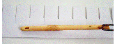
Figure 1: Fragment of a urinary catheter taken from
an inpatient in the intensive care unit-Sidi Bel
Abbes University Hospital - Algeria. Use of sterile graduated rule.
Catheters can have a large variety of sizes,
constituents and types. Clinician has to keep in mind numerous factors, e.g.,
medical necessity, expected time of use, individual choice and the infection
risks involved [15, 16].
For these reasons, we suggest for greater precision,
that the results of the CFU / mL or cell / mL evaluation be supplemented by the
unit length. Conversely, the neglect of these last two parameters, the length
of the removed part of the catheter and the diameter thereof, may lead to
visibly erroneous results as to the microbial load of the catheter removed;
therefore, the significance level will be incorrect.
1. Conclusion
The diagnosis of catheter infectivity involves several
parameters related to the clinical information of the patient and the microbial
presence on the catheter after culturing. The catheter infectivity type is
related to the level of significance. Nevertheless, differences in dimensions
do exist between several kinds of catheters. For this, future studies should
contemplate this parameter to properly evaluate
the microbial load.
2. Acknowledgment
We would like to thank Dr. Asma
Kebiri for reading the English version of this
article.
3. Conflict of interest statement
We certify that there
is no conflict of interest with any financial organization in the subject
matter or materials discussed in this manuscript.
4. Authors’ biography
No Biography
5. References
[1]. Maki, DG, Jarretf F, SarafinHW. 1977.
A semi quantitative
culture method for identification of
catheter-related infection in
the burn patient.
J. Surg. Res.
22(5): 513-520. https://doi.org/10.1016/0022-4804(77)90034-8
[2]. Cleri DJ, Corrado ML, Seligman SJ. 1980. Quantitative Culture of Intravenous
Catheters and Other Intravascular Inserts. J. Infect. Dis. 141(6): 781-786. https://doi.org/10.1093/infdis/141.6.781
[3]. Brun-Buisson C, Abrouk F, Legrand P, Huet Y, Larabi S, Rapin M.
1987.Diagnosis of central venous
catheter-related sepsis: critical
level of quantitative
tip cultures. Arch.
Intern. Med. 147(5): 873-877. https://doi.org/10.1001/archinte.147.5.873
[4]. Seddiki SML, Boucherit-Otmani Z, Mahdad YM, Bendahmane AF,
Kunkel D. 2018.Proposition of an
appropriate technique to diagnose catheters fungal infectivities.JKSUS.
30(3) : 400-403. https://doi.org/10.1016/j.jksus.2018.04.012
[5].
Goldman, Emanuel; Green,
Lorrence
H 2008. Practical Handbook
of Microbiology, Google eBook, 2nd
ed., CRC Press, Taylor and Francis Group. p. 864.ISBN
978-0-8493-9365-5.Retrieved 2018-10-16.
[6].
Carrière C, Marchandin H.
2001. Infections liées
aux cathéters veineux
centraux : diagnostic et
définitions. Néphrologie. 22(8): 433-437.
[7]. Mermel LA, Farr BM, Sheretz RJ, Raad II, O'Grady N, Harris
JS, Craven DE. 2001.
Guidelines for the management of intravascular catheter-related infections. Clin. Infect. Dis. 32(9): 1249-1272. https://doi.org/10.1086/320001
[8]. Seddiki SML, Otmani-Boucherit Z, Boucherit K, Badsi-Amir S, Taleb M, Kunkel D.
2013. Assessment of the
types of catheter
infectivity caused by
Candida species and
their biofilm formation.First study
in an intensive
care unit in
Algeria. Int. J.
Gen. Med. 6,
1-7. https://doi.org/10.2147/IJGM.S38065
[9]. Ryan JA, Abel RM, Abbott WM, HopkinsCC,
Chesney TM, Colley R, Phillips K, Fischer JE.1974. Catheter
complications in total
parenteral nutrition.A prospective
study of 200 consecutive patients. N. Engl. J. Med. 290(14) : 757-761. https://doi.org/10.1056/NEJM197404042901401
[10]. Domart Y., Hoen B, Leport C,
Cartier F, le
Groupe de travail
sur les infections cardiovasculaires. 1991.
Traitement curatif des
infections sur cathéter
veineux central en fonction
du germe, de
la situation clinique
et du type
de cathéter (cathéter
en place ou
après ablation). Propositions, limites.Nutr.Clin.Métabol.
5(2) 95-104. https://doi.org/10.1016/S0985-0562(05)80116-5
[11]. Brun-Buisson C. 1994. Analyse critique des
méthodes diagnostiques d'infection liée au cathéter sur matériel enlevé.
Réan.Urg.3 (3 bis) 343-346.https://doi.org/10.1016/S1164-6756(05)80726-1
[12]. Rello J, Cell P, Prats G. 1991. Laboratory
diagnosis of catheter-related bacteremia. Scand. J. Infect. Dis. 23(5):
583-588. https://doi.org/10.3109/00365549109105182
[13]. Snydman DR, Gorbea HF, Pober BR, Majka JA, Murray SA, Perry LK. 1982. Predictive
value of
surveillance skin cultures
in total parenteral nutrition-related infections.
Lancet. 18(2):1385-1388. https://doi.org/10.1016/S0140-6736(82)91281-8
[14]. Collignon PJ, Seal
N, Pearson IY,
Woods WP, Munro
R, Sorrell TC.
1986. Is semi-quantitative culture of central vein
catheter tips useful in the diagnostic of catheter associated bacteremia? J. Clin. Microbiol. 24(4): 532-535.
[15]. Meddings, J., Rogers,
M. A., Krein, S. L., Fakih, M. G., Olmsted, R. N.,
& Saint, S. 2013. Reducing unnecessary urinary catheter use and other
strategies to prevent catheter-associated urinary tract
infection: an integrative
review.BMJ
quality & safety,
23(4), 277-89. https://doi.org/10.1136/bmjqs-2012-001774
[16]. Cai Z, Chattopadhyay
N, Liu WJ, Chan C, Pignol JP, Reilly RM 2011.
Optimized digital counting colonies of clonogenic
assays using ImageJ software
and customized macros: comparison with
manual counting. Int
J Radiat Biol. 87
(11): 1135-46. https://doi.org/10.3109/09553002.2011.622033